Thinking your doctor is an artist doesn’t make it so: Eye Diagnostics vs. Hand Drawing in the 21st Century? Your Choice!

2 03 2014

Slide1Ages ago, doctors didn’t have the ability to image structures so they relied heavily on careful detailed written description to accompany hand drawn diagrams to monitor their patients.  Sounds archaic doesn’t it?  Living in 2014, we now have a breadth of tools that can help us see structures in the body that we can’t even see with the naked eye – if given the choice of a hand drawn picture with written notes versus an advanced diagnostic image of the organ in question, which would you prefer to have done?  What if it could save your vision or your life?  With current electronic medical record keeping, there often isn’t an option to draw so a detailed description is usually the only option we doctors have to document your condition.  Depending on the space in the patient record that 1 or 2 sentence description of the part of the body is used to remind the doctor a year or two later what she or he saw at the previous visit and make clinical decisions on the health status of that particular part of your body.  Does that worry you?

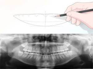
Can you imagine your dentist NOT taking an x-ray to monitor your or your child’s oral health and development?  Relying on written word to describe the anatomy to make clinical decisions in 2014?  Considering everyone with a smartphone is capturing their living history by snapping away at everything from the breakfast they eat to the party last night, it seems like our memory is getting worse or at least we are less willing to rely on them.teeth_xray
The organ I look after is the eye, and there are several areas of the eye that require careful and detailed attention to changes in size, thickness, elevation, artery angles, reflectivity, etc. (I could go on).  With the advances in technology, imaging the eye is now possible such that we can even see  through the tissues and assess what is not visible under a microscope or to the naked eye.  We can now track discrete changes down to a fraction of a hair using digital quality images and video so that the slightest change becomes part of the patient record for generations to come.   We can invert, flip, and virtually turn the eye inside out in order to assess potential holes, bleeding, retinal detachments, etc.  Gone are the days where we to rely on deciphering our written notes from a year ago to monitor for change – or are they?

Depending on where you live, these types of images are not covered by government insurance plans and even some private plans.  Their lack of coverage however doesn’t make them any less relevant to your ocular health.  In fact it is a well accepted science that imaging the optic nerve, retina and other ocular structures provides earlier diagnosis and better overall management. fundus_drawingWith an image set, your eye doctor can review areas over and over with scrutiny without having to subject the patient to holding their eye open with a bright light being blasted at them.  It takes away the issue of patient movement while examining the eye, which makes it extremely difficult to provide an accurate and efficient assessment.  For children this is particularly helpful.  With this combined information of an image set and your doctors microscopic evaluation, this provides you with the most comprehensive  assessment using 21st century technology.

What is astounding to me is that despite what we know about the relevance and importance of the diagnostic eye imaging, there are still  some patients that don’t seem to  understand its utility and choose to not have it done – like opting out of a warranty at Best Buy.   With all advances in technology, very few have failed to adopt;  do you still use a VCR or an 8-track tape player to watch your movies or listen to music? If my mechanic requests that I run a diagnostic on my car, I generally rely on her or his professional judgement to decide on having that test done.  I can’t think of the last time I said “no’ to a dental x-ray.  In general I rely heavily on the experience of my service providers to recommend what is best.

My patients are educated thoroughly on why I make this type of diagnostic choice in order to provide the most thorough of eye care.  Prevention and wellness are as much a part of health care as the treatment we provide for what ails you.  In fact, identifying earlier and intervening before major damage has set in can save your vision for years to come.  Diabetes for example doesn’t have to cause blindness. With regular comprehensive eye examinations and retinal diagnostics, we can actually prevent this disease from taking your vision away.  Countries like the UK that have a national program employing such diagnostics are actually seeing a stabilization in the number of diabetes related blindness after almost 10 years of implementation.

So why are we opting out in North America of retinal diagnostics despite what eye doctors know to be the best way to preserve your most precious of senses?  Why don’t we provide the most comprehensive care on 100% of patients?  Have we commoditized eye care and the value of our doctor’s extensive clinical training and experience?  Do patients value vision the same way we negotiate for a car?  For the sake of all of our eyes – I hope not.

In good health,

Dr. Richard Maharaj OD, FAAO

Cinical Director,

eyeLABS Optometry and Center for Ocular Surface Disease

www.eyelabs.ca

twitter: @eyelabsinc

info@eyelabs.ca


Do you suffer from acne? Bumps on your eyelids can be a sign

21 01 2014

zitDo you suffer with zits or chronic acne on your face or elsewhere on your body?  Are you obsessed with having and maintaining clear skin?  If you don’t already know, a zit or a pimple on your skin happens when the oils in your pores are trapped, built up and harden.  Your ‘pores’ on your skin are actually called sebaceous glands and are oil producing glands that give your skin the glowing complexion.  Irregular production and secretion of these oils can be caused by dirt from your hands and makeup and lack of proper cleaning of your skin from dead tissue or built up debris.  When these glands, or pores, become obstructed the skin tissue becomes inflamed turning red swollen and sometimes painful.  This inflammation is the body’s response to local trauma.  Now this isn’t the trauma caused from a hit or a poke, but microtrauma induced by the trapped oil and debris pushing on the surrounding wall of the gland and skin.  Some people with certain skin types have a strong inflammatory response which in biology is known as the triple response (of Lewis) which consists of:

  1. Red spot: due to small capillary dilatation
  2. Flare: redness in the surrounding area due to arteriolar dilatation
  3. Wheal: due to leakage of fluid from capillaries and venules

Those with a stronger response will be more likely to have stubborn acne that has limited responses to commercial products.  These cases usually require a skin specialist (dermatologist) to treat it medically.  People with eczema, asthma, psoriasis and rosacea (to name a few) are known to have a hypersensitive triple response.

meibglandNow that you understand what causes a pimple, you should also know that those sebaceous glands found on your skin are also found in your eyelid and are slightly modified to secrete oils for the eye’s surface.  Each eyelid contains 20-40 of these glands and have a very specialized function.  These glands are called meibomian glands and react the same way the pores on your skin react to debris, dirt, bacteria and general trauma.  What’s different about this area of the body however is that your eyelid is constantly moving and wiping away environmental debris from the ocular surface.  If you are a contact lens wearer, then it is also rubbing the plastic of the contact as well causing friction on the inner eyelid.  Considering what we know about trauma and what it does to our glands, imagine that this microtrauma happens every time you blink, every time you apply make up, every time you wear a contact lens and every time your eye is exposed to environmental debris.  That is pretty much every minute of every day!   The average human blinks seven to ten thousand times a day and for those people that have a heightened inflammatory response are more susceptible to the meibomian glands becoming obstructed which can lead to an eyelid pimple otherwise known as a stye.  These styes can get very large if untreated and become chalazion or an eyelid cyst which can in some cases require surgery.  Interestingly, a stye only happens when the blockage has gotten large enough to become visible to the human eye.  The blockage usually starts long before it becomes visible externally.

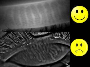
Unfortunately though, these meibomian glands are few and once blocked or inflamed can quickly become dysfunctional and die off. happysad The eyelid surface is uniquely situated close to the eyelash follicles.  Naturally occurring bacteria often accumulate in high numbers in this area due to built up dead skin, makeup and environmental debris.  This surface cannot be cleaned by commercial products with the precision required without causing harm to the eye (cleanser in the eye, removal pads can abrade the cornea, etc.).  Also the inner eyelid tissue known as the wiper is too sensitive to touch or use retail cleaning agents on without causing pain and or damage.  Basically this tissue is never really cleaned and these glands are under constant burden.

Your eyelid glands are just like your skin glands and need clinical attention to keep them functional and prevent meibomian gland dysfunction which causes up to 86% of dry eye disease patients.  In many cases it is a preventable condition, but only recently has clinical periocular hygiene emerged as a new effective maintenance treatment.  Using special dyes and instruments, the eyelid surface and glands can be treated comfortably in your eye doctor’s office.   New advances in this area allow even the most hardened blockages to be melted using prescribed heat and pressure simultaneously (LipiFlow thermal pulsation) in non-surgical painless procedure.

Bumps on your eyelid are exactly the same as pimples on your face, however the consequence of not treating it or preventing it are potentially vision threatening.  If left untreated eventual gland death can occur decreasing the stability and vitality of your tearfilm.  This puts the entire surface of the eye at risk for infection, chronic inflammation and even scarring of the conjunctiva or cornea.

Experts from around the world have now recognized meibomian gland dysfunction as being perhaps the most pervasive cause of dry eye disease around the planet.  These tiny delicate pores have a huge impact on our visual health.  Talk to your eye doctor about your glands and be proactive.  Having dedicated my career to the ocular surface and spending the last 2 years specifically developing treatments in this area, my advice is:  don’t let the symptoms start!

In good health,

Dr. Richard Maharaj OD, FAAO

Cinical Director,

eyeLABS Optometry and Center for Ocular Surface Disease

www.eyelabs.ca

twitter: @eyelabsinc

info@eyelabs.ca


20/20 isn’t everything: See why every child MUST have a comprehensive eye exam to prepare for school

17 10 2013

Dr. Richard Maharaj and Dr. Chris Schell demonstrate some common vision problems that children struggle with everyday in the classroom. A very important message on why comprehensive eye examinations (not a vision screening) must happen for each child prior to (or at the very least, as soon as possible) school age. Vision screenings are well-intentioned, but as shown in the video, may miss these subtle diagnoses and give parents a false sense of security. Their little eyes are starting a 12+ year marathon which will serve the basis of learning.  You wouldn’t run a real marathon without preparing your muscles and endurance would you?  Why do we assume newly developing eyes should be treated with less concern or preparation.  See a Doctor of Optometry for a comprehensive eye examination.


Dr. Chris Schell discusses Ontario’s Eye See Eye Learn Program on CTV news

28 08 2013

Dr. Chris Schell is featured on CTV Barrie discussing the Eye See Eye Learn program in Ontario. He discusses the importance of children’s eye examinations for school aged children. Dr. Schell has a special interest in Vision Therapy and Pediatric Optometry.


A Child’s Vision: How important is it to you

19 08 2013

Only 14% of children under 6 in Canada have had a comprehensive eye exam yet 80% of learning is visual. In preparation for a 12+ cycle of education is this how do we equip our kids? We should expect more. Many common learning disabilities have been shown to be hidden undiagnosed visual dysfunction. Schedule your child’s eye exam today!


Be Proactive on Reducing Wrinkles – add comfort to your eyes!

25 07 2013

“Epidermal atrophy and structural changes observed in the Dermal-Epidermal Junction with aging may be, by some extent, related to daily and repetitive skin deformations all along the life span”

Skin Res Technol. 2013 Jun 25

Translation:  Wrinkles and the aging skin are related to constant stretching and relaxing of the skin.  This may sound like common sense, yet patients everywhere still engage in life behaviours that are known to hasten wrinkle formation.   Smoking, tanning, high sugar diets all are big contributors to the aging skin but while changing your habits may take some work, why not eliminate some factors that are out of your everyday control.

crowsfeet

Squinting your eyes – a very common ocular reflex to external factors such as bright light, blurry vision and ocular discomfort.  Because it’s a reflex it is not considered a bad habit, but rather a mechanism your body employs to protect the ocular surface and or enhance vision when stimulated by the above factors.  Here are some very easy steps to reduce your stimuli to squint:

  1. Wear corrective eye wear when needed – this is by far the easiest step to take to avoid squinting.  It has the added benefit of clearing your visual world.
  2. Wear your sunglasses – UV exposure worsens this cause of squint because it can accelerate the aging skin.  If you’ve ever lost your sunglasses and spent even a lunch hour without them, you’ll likely have a headache all afternoon.
  3. Treat your ocular surface keeping it wet and comfortable – Dry Eye Disease (DED) is a leading cause of eye discomfort.  When left untreated the natural course of this disease will worsen leading to further eye muscle contraction.

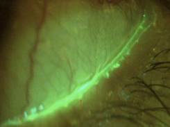
The treatment options for dry eye have changed dramatically as experts world wide agree that meibomian (oil) gland dysfunction (MGD) may be the leading cause of dry eye in the world (TFOS 2011).  Knowing the mechanism behind this condition allows it’s treatment to be more targeted and therefore more effective.  Each of the four eyelids are home to the 20 to 40 oil glands and optimal functioning is achieved when the glands are absent of any obstruction and the lid surface is properly cleared of debris and devitalized epithelium.  The eyelid margin (the area behind your eyelashes towards the eye) is the only area of our bodies that doesn’t get cleaned regularly or thoroughly and because of its proximity to the cornea is a dangerous place to try to clean at home with q-tips, brushes or wipes.  Corneal scratches can become infected and are extremely painful.  Also, over the counter cleansers are still insufficient to removed devitalized skin and debris which can only be seen under microscopic examination with ophthalmic vital dye staining.LOM_mgo

eyeLABS Center for Ocular Surface Disease offers an array of services to treat and maintain your glands and lids for optimal functioning:

  • Lid Margin Debridement – and exfoliative process for the eyelid surface
  • BlephEx – a microdermal exfoliation of the eyelid surfaces and eyelash roots
  • LipiFlow Theramal Pulsation – the only FDA approved treatment for MGD.
  • Topical and Oral Medicines – although we focus on enhancing your body’s natural functioning there are cases that require specific medicine to treat underlying inflammation and/or infection

In this way, we can limit your stimulus to squint and help you widen those eyes and lose the wrinkles!  Instead of spending countless dollars on Botox and fillers, be proactive and prevent unnecessary “epidermal atrophy,” also known as wrinkles!

Dr. Richard Maharaj OD, FAAO

Director of Optometry,

eyeLABS Center for Ocular Surface Disease

www.eyelabs.ca

twitter: @eyelabsinc

rmaharaj@eyelabs.ca


Leaving a Legacy – The passing of Dr. William Samis

13 06 2013

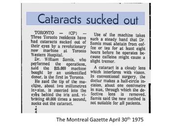
The Montreal Gazette April 30th 1975On June 12th 2013, we lost a very good man and a groundbreaking surgeon – Dr. William Samis.  In 1975, he became  the first ophthalmologist in Ontario to perform phacoemulsification surgery for cataract patients and one of the first in the world to adopt this then groundbreaking technique and associated lens implants.  Indeed a visionary, he cultivated an environment which bred future leaders in the his field.  I joined his practice (partnered with Dr. Steve Arshinoff MD) in 2005 and was touched by his generosity as a human being, his style and sense of humour.  Unbeknownst to him, I absorbed a great deal from this man as an eye doctor and as a person through his words of wisdom and through the eyes of his patients, many of which I adopted after he stopped seeing patients from being ill 2 years ago.  Each an everyone would comment on his candor and caliber as a friend first, and a physician second and every last one would sing his praises as a surgeon decades after having had cataract surgery by his hands.

I like to think I’ve learned even an ounce of what this man had taught me and the many surgeons who walked through his doors or were privileged enough to have learned their surgical skills as his resident.

I have a hard time accepting that he is gone, however I have a harder time believing that for 6 years I was able to work closely with a true game changer – an individual who literally transformed modern eye surgery to make it faster, safer while producing better outcomes.    Some people wish to win a lottery – I believe I did.

My deepest sympathies go out to his family, his wife Jane, and dear friend and colleague Dr. Steve Arshinoff.  We were all in the presence of greatness.

RM


Eye on Eyes: Irlen Syndrome, VEGF inibitors and a TED talk to guide better science

25 05 2013

Dear Readers:

This edition is a shift from the regular.  In conversations with colleagues and optometry forums I’ve decided to share some of the current conversations being had in optometry, ophthalmology and in medicine.  In this edition, I discuss Alberta’s Bill 204 which is a pending act that brings vision therapy and behavioural optometry into the spotlight.   Retina online published an interesting look at the incidence of nonfatal MI’s and how VEGF inhibitors may increase patient risk.  Finally, for those that know me know my frustration with faulty science and ‘counterknowledge,’  I share a video and encourage practitioners of all disciplines to look at Ben Goldacre’s TED talk on the subject.

In good health,

RM

Irlen Syndrome Testing Act Bill 204 in Alberta Legislature Second Reading

My friend and colleague Dr. Chris Schell (http://www.optometristbarrie.ca/) brought up an interesting piece of legislation in the Alberta government with respect to early learners eye screening.  This bill, also known as the Irlen Syndrome Testing Act, will call for School Boards to make available screeners for Irlen syndrome (including scotopic sensitivity syndrome and visual disturbance syndrome).

Section 3 is quoted below:

3(1) A teacher who recognizes symptoms of Irlen Syndrome in a student must recommend to the parents or guardian of the student that he or she be tested for Irlen Syndrome.

(2) Upon receiving written consent from the parent or guardian of a student, that student must be tested by a screener who is made available by a board under section 2.

(3) If a screener determines that the student may have Irlen Syndrome, the screener must advise the parent or guardian and indicate what corrective measures may be undertaken and if additional testing is required.

This bill is certainly an interesting step in the broader acceptance of vision therapy, however the increasing information on Irlen Syndrome  calls into question the syndrome itself.  Schiemann et al. has demonstrated that there is significant overlap in symptoms of Irlen syndrome patients and the majority (95%) have unresolved binocular vision and refractive anomalies (J Am Optom Assoc, 1990).  The ‘success’ of Irlen filters may be in fact indicative of  more specific binocular vision syndrome and the improvement by filtered lenses may be more of a provocative test than a treatment in my opinion.  So although the bill is well intentioned, I fear the shoulder’s that it is being rested on.

Click here to See Bill 204

Myocardial Infarctions following Intravitreal VEGF Inhibition
(Retina Online Vol 9 No. 5)

The authors of the following study sought to determine the risk of thromboembolic and gastrointestinal bleeding events in the 12 months after injections of bevacizumab or ranibizumab compared with photodynamic therapy (PDT) and a nontreated community sample.

They examined hospital and death records for 1,267 patients treated with vascular endothelial growth factor (VEGF) inhibitor and 399 patients treated with photodynamic therapy (PDT) attending Western Australian eye clinics from 2002 to 2008, and 1,763 community controls, aged ≥50 years. They also analyzed hospital records from 1995 to 2009 for history of myocardial infarction (MI), stroke and gastrointestinal bleeding before treatment. They searched records for evidence of these events in the 12 months after treatment.

The 12-month MI rate was higher for VEGF inhibitor patients than PDT and the community group (1.9/100 vs. 0.8 and 0.7, respectively), the authors reported. They observed no differences between patients treated with bevacizumab and ranibizumab, and the noted that the adjusted MI rate was 2.3 times greater than the community group (95% confidence interval, 1.2–4.5) and PDT rate (95% confidence interval, 0.7–7.7). The 12-month MI risk did not increase with the number of injections administered (hazard ratio, 0.9; 95% confidence interval, 0.5–1.5). Stroke and gastrointestinal bleeding did not differ between any exposure groups.

Although all of the adverse events examined were rare, patients treated with VEGF inhibitors were significantly more likely to experience fatal or nonfatal MI than the community group, the study authors determined. This increased risk may be related to the underlying age-related macular degeneration or vascular endothelial growth factor inhibitor use itself.

Source: Kemp A, Preen DB, Morlet N, et al. Myocardial infarction after intravitreal vascular endothelial growth factor inhibitors: a whole population study. Retina. 2013;33(5):920–927.

 Bad Science – a TED talk on Practicing Mindful  Medicine and Steering Clear of Counter-science

“It was the MMR story that finally made me crack,” begins the Bad Science manifesto, referring to the sensationalized — and now-refuted — link between vaccines and autism. With that sentence Ben Goldacre fired the starting shot of a crusade waged from the pages of The Guardian from 2003 to 2011, on an addictive twitter feed, and in bestselling books, including Bad Science and his latest, Bad Pharma, which puts the $600 billion global pharmaceutical industry under the microscope. What he reveals is a fascinating, terrifying mess.

Click the image to view


TEDBENGOLDACRE


Just another BRVO?

21 05 2013

A 39 year-old male of South Asian decent reported to the clinic reporting blurry vision in his right eye starting 3-4 days previous.  Vision measured OD 20/50 OS 20/20 (uncorrected – this patients was previously 20/20 OD and OS).  Patient history revealed self reported ‘mild’ hypertension which was not medically managed nor had it been indicated in previous visits with his primary care physician.   IOP was 17 mmHg OD/OS and pupils were normal.  Flurescein angiography study showed no ischemia, however a conservative approach was taken to monitor the macula edema for resolve rather then consider anti-VEGF or therapeutic laser options at the time.

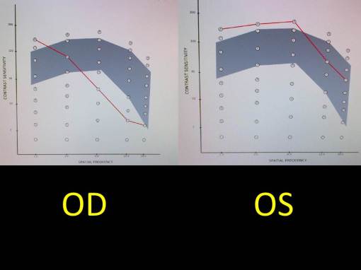
Fundus photo and OCT are shown below OD as well as contrast sensitivity testing.

ODBRVOwMEThe BRVO and to a lesser extent the macular edema is obvious on fundus examination.  On closer inspection though another area off inferiorly and nasal to the disc shows vascular retinopathy in the form of a cotton wool spot and nerve fibre hemorrhages.  This is not associated with the BRVO and signals the chronicity of his systemic hypertension which resulted in an interesting turn of events for this otherwise carefree individual.

Macular edema2ndBRVO

The OCT of the macula  clearly shows the cystic edema associated with this inferior BRVO.  Because of its inferior location this fluid will likely drain away from the macula and be reabsorbed in natural course.

Notice the difference in CS vs. SF curve.  This test was done for monitoring purposes only – it was not a diagnostic test.  It was needed to monitor the quality of his vision in this case as the fluid reduced at his macula.  The retinal surgeon and myself  will rely on this in addition to his objective findings downstream to help in guiding treatment options in the event of poor resolution of edema.

ContrastSensitivityBRVO

Follow up and Discussion:

From a bird’s eye view, I would say this case is worthy of follow up but not a unique set of findings.  What followed was however very interesting.  I examined this patient on a Thursday.  I called his family doctor but was unable to speak to her.  I sent a report and advised to follow up on his hypertension at next visit which turned out to be the next day.   This patient was very proactive and I would argue, his proactive nature helped to save his own life.   Serial BP testing demonstrated an avg BP of 180/160 and the final measurement of the day climbed past 200/180 sending this patient to the emergency room at the local hospital.  He was admitted to CCU over the next 3 days in an  attempt to urgently reduce his risk of stroking out and to steer this patient away from certain death.  After an intense 3 days his BP had come down to 130/85 in response to his medical therapy and it was maintaining.  The patient reported  that his multiple daily headaches had stopped and his quality of sleep was markedly improved.

I am following him regularly until complete resolution, however I believe there is a lesson learned here.

Better communication – For years, this  man was told he was ‘fine’ but had mild hypertension.  His cultural background supported a holistic approach to managing one’s body.  Through mind and inward discipline he was confident that he was in good health.   He hadn’t followed up regarding the ‘mild’ hypertension for at least 2 years because it wasn’t a concern for his doctor so why should it be a concern for him?   In fairness, there was no way to predict that he would suffer from hypertensive crisis years later so the ‘see your doctor when there’s a problem’ approach seemed prudent here.

My communication as his eye doctor to his family doctor was difficult and I haven’t yet received a report on this potentially critical patient whom I referred for urgent care.    To be clear I don’t believe this to be a problem with this doctor specifically but with the system at large.  This is what I believe needs to change.  How we relay information to each other; whether it is two teenagers tweeting their breakup because of broken communication or a doctor to her or his patient or physicians reporting over the care of a patient we are all too busy, too slavish to outdated protocol, too pre-occupied with all about us to listen.

For this lucky young man, I suspect he is listening to his body more than ever.  He told me he and his wife are finally going to take their honeymoon which is years overdue and will slow things down a little at work.  For the record I am biased to the sense of sight,  but I think we could all do a little better to listen to the world around and within us.

In good health,

Dr. Richard Maharaj OD, FAAO

Director of Optometry,

eyeLABS Inc.

www.eyelabs.ca

twitter: @eyelabsinc

rmaharaj@eyelabs.ca


25.8 Million US with Diabetes vs. 23 Million with Dry Eye Disease

16 05 2013

In June 2012, eyeLABS center for Ocular Surface Disease became the first optometry clinic in Canada to offer LipiFlow Thermal pulsation treatment – the only FDA approved treatment for evaporative dry eye.  eyeLABS is a unique facility because we don’t sell glasses – we manage the ocular surface of patients far and wide and we do so with an array of options that allows for a customized approach to managing a very complex disease – Dry Eye Disease.

eyeLABS year 1 clinical results:

Looking  just over 100 eyes treated using a combination of LipiFlow, lid management techniques, and medicine we have achieved a 90% rate of significant improvement in clinical signs and patient symptoms, 8% with mild to moderate improvement and 2% minimal to no change.  This was defined in our clinical study by validated patient symptom scores, gland scores, corneal staining, tear film break up time and visual acuity.  As a result we continue to educate as many physicians and patients as possible about the shift in approach of managing dry eye from exclusively cornea to the eyelids and glands.

Prevalence of DED vs. Diabetes

According to NDEP (National Diabetes Education Program) there are 25.8 million Americans living with diabetes.  According to a Marketscope 2011 Comprehensive Report on the Global Dry Eye Products, there are 23 million patients living with Dry Eye Disease.  Of course the comparison in the impact of two diseases on the body isn’t fair, but the impact on quality of life (QoL) is eerily similar.  A 2012 study examining 87 dry eye patients and 71 healthy volunteers found that vision-related QoL in dry eye patients was impaired and was correlated with anxiety and depression( Li, M Invest Ophthalmol Vis Sci. 2012 Aug 17).  Countless studies examining QoL within the diabetic subset show strong correlations to anxiety and depression due to the daily burden of medicine, monitoring and management.

Dry eye is too often dismissed by physicians as insignificant and ‘not as important’ as other ocular ailments like cataracts or glaucoma and patients have become embarrassed to mention it.  In fact, when eyeLABS Dry Eye Clinic patients were surveyed, the most common reason for not talking about dry eye with other doctor was embarrassment.  Embarrassment that their doctor wouldn’t think it was important.  Interestingly all patients reported social anxiety about the cosmetic appearance of their red eyes to colleagues, family, friends etc. and had sought out medical attention to treat the anxiety as a result.  eyepicture

The social impact of these two diseases are  far-reaching.  Given that the prevalence of the disease is near equivalent why is it that dry eye is swept under the rug?  It is possible that medical options of dry eye have been limited and expert agreement on the cause is divided has resulted in doctor’s complacency towards this growing epidemic.

Being in the position that I’m in seeing dry eye and ocular surface day in day out I can say that this condition does deserve attention and undivided attention at that.  My patients are physicians, teachers, celebrities, pilots, mothers, fathers and and they have all opened up about the anxiety that dry eye has caused them.  One might assume that those listed above are ‘professionals’ and would never leave the disease to take over their lives but many  patients have even considered suicide prior to having treatment because of how limited their lives had become.   For the first time they have had relief and I feel grateful for the opportunity to change their lives.

When one considers the impact of a disease on mental health, the mental health should be then considered a co-morbidity.  Diabetes, Dry Eye Disease and many other ailments all have the common denominator on reduction in quality of life and an increase in depressive and anxious tendencies.  Almost 50 million Americans share this common denominator between diabetes and dry eye, but those with dry eye don’t have nearly as many resources for dealing with their condition.  Considering the success I’ve had with my patients in this last year, I submit that we can do a better.

In good health,

Dr. Richard Maharaj OD, FAAO

Director of Optometry,

eyeLABS Inc.

www.eyelabs.ca

twitter: @eyelabsinc

rmaharaj@eyelabs.ca