Thinking your doctor is an artist doesn’t make it so: Eye Diagnostics vs. Hand Drawing in the 21st Century? Your Choice!

2 03 2014

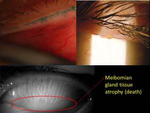
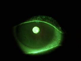
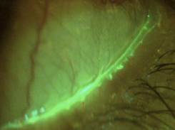
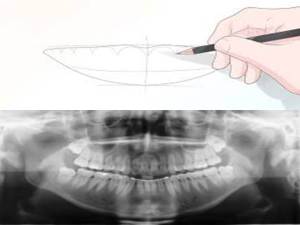
Slide1Ages ago, doctors didn’t have the ability to image structures so they relied heavily on careful detailed written description to accompany hand drawn diagrams to monitor their patients.  Sounds archaic doesn’t it?  Living in 2014, we now have a breadth of tools that can help us see structures in the body that we can’t even see with the naked eye – if given the choice of a hand drawn picture with written notes versus an advanced diagnostic image of the organ in question, which would you prefer to have done?  What if it could save your vision or your life?  With current electronic medical record keeping, there often isn’t an option to draw so a detailed description is usually the only option we doctors have to document your condition.  Depending on the space in the patient record that 1 or 2 sentence description of the part of the body is used to remind the doctor a year or two later what she or he saw at the previous visit and make clinical decisions on the health status of that particular part of your body.  Does that worry you?

Can you imagine your dentist NOT taking an x-ray to monitor your or your child’s oral health and development?  Relying on written word to describe the anatomy to make clinical decisions in 2014?  Considering everyone with a smartphone is capturing their living history by snapping away at everything from the breakfast they eat to the party last night, it seems like our memory is getting worse or at least we are less willing to rely on them.teeth_xray
The organ I look after is the eye, and there are several areas of the eye that require careful and detailed attention to changes in size, thickness, elevation, artery angles, reflectivity, etc. (I could go on).  With the advances in technology, imaging the eye is now possible such that we can even see  through the tissues and assess what is not visible under a microscope or to the naked eye.  We can now track discrete changes down to a fraction of a hair using digital quality images and video so that the slightest change becomes part of the patient record for generations to come.   We can invert, flip, and virtually turn the eye inside out in order to assess potential holes, bleeding, retinal detachments, etc.  Gone are the days where we to rely on deciphering our written notes from a year ago to monitor for change – or are they?

Depending on where you live, these types of images are not covered by government insurance plans and even some private plans.  Their lack of coverage however doesn’t make them any less relevant to your ocular health.  In fact it is a well accepted science that imaging the optic nerve, retina and other ocular structures provides earlier diagnosis and better overall management. fundus_drawingWith an image set, your eye doctor can review areas over and over with scrutiny without having to subject the patient to holding their eye open with a bright light being blasted at them.  It takes away the issue of patient movement while examining the eye, which makes it extremely difficult to provide an accurate and efficient assessment.  For children this is particularly helpful.  With this combined information of an image set and your doctors microscopic evaluation, this provides you with the most comprehensive  assessment using 21st century technology.

What is astounding to me is that despite what we know about the relevance and importance of the diagnostic eye imaging, there are still  some patients that don’t seem to  understand its utility and choose to not have it done – like opting out of a warranty at Best Buy.   With all advances in technology, very few have failed to adopt;  do you still use a VCR or an 8-track tape player to watch your movies or listen to music? If my mechanic requests that I run a diagnostic on my car, I generally rely on her or his professional judgement to decide on having that test done.  I can’t think of the last time I said “no’ to a dental x-ray.  In general I rely heavily on the experience of my service providers to recommend what is best.

My patients are educated thoroughly on why I make this type of diagnostic choice in order to provide the most thorough of eye care.  Prevention and wellness are as much a part of health care as the treatment we provide for what ails you.  In fact, identifying earlier and intervening before major damage has set in can save your vision for years to come.  Diabetes for example doesn’t have to cause blindness. With regular comprehensive eye examinations and retinal diagnostics, we can actually prevent this disease from taking your vision away.  Countries like the UK that have a national program employing such diagnostics are actually seeing a stabilization in the number of diabetes related blindness after almost 10 years of implementation.

So why are we opting out in North America of retinal diagnostics despite what eye doctors know to be the best way to preserve your most precious of senses?  Why don’t we provide the most comprehensive care on 100% of patients?  Have we commoditized eye care and the value of our doctor’s extensive clinical training and experience?  Do patients value vision the same way we negotiate for a car?  For the sake of all of our eyes – I hope not.

In good health,

Dr. Richard Maharaj OD, FAAO

Cinical Director,

eyeLABS Optometry and Center for Ocular Surface Disease

www.eyelabs.ca

twitter: @eyelabsinc

info@eyelabs.ca


Learning from failures – My top 3 dry eye treatment mishaps

22 01 2014

Having practiced for over 10 years now, I’ve had the most success and most impact on my dry eye patients in the last 2 years.  I just celebrated a milestone of having helped 200 patients achieve a level of clinical stability and symptomatic relief of their chronic dry eye disease.  I attribute that success to the breadth of clinical knowledge and research that I’ve put into building the dry eye clinic in addition to the dramatic increased volume of peer reviewed science directed towards the study of dry eye disease and its causes. More than anything else, however I attribute my patients success to the cases that failed initial treatment.  A good scientist’s successes are shouldered by his or her failures.

In this brief article I want to share from my dry eye cases that responded poorly or not at all to therapy.  It’s funny but 3 specific cases always come to mind when I think about this subject.  One case involves Lyme disease, another was a male with ‘borderline-normal’ testosterone and the last was a case of ‘he said she said’.

corneaesthesia1) Patient S.A. had been battling a diagnosis of Lyme disease when she presented to my office and all clinical signs pointed to MGD.  Meibography showed mild truncation but nothing more than I had seen in my most successful cases, and they certainly had viable expression on forced palpation.  Despite effective clearing of obstruction using LipiFlow (confirmed on post meibography) and improved ocular surface staining, she remained with mild improvement in meibum expression.  Her symptoms, as often observed with dry eye disease failed to match the improved clinical picture.   Systemically she was also not improving which I attributed to her lack of improvement.  However on closer inspection corneal sensitivity pre-treatment and 6 months later had increased.  I had assumed that initial testing was basal and normal, however it was more likely that this patient had experienced hypoaesthesia on presentation and treatment resulted in increased surface threshold sensitivity – a return to normal feeling if you will.  Lesson:  Longstanding cases of DED with and without systemic involvement will at some point undergo neural upregulation (or dysregulation) which can and will confuse the clinical picture.  I’ve learned from this that staying the course in the interest of decreasing inflammation is prudent, despite a failing symptomatic picture.  Sometimes feeling anything is better then feeling nothing at all!

nomgd2) Patient M.H.  had been to 3 corneal specialists in the previous 5 years.  He had been on various doses of doxycycline, restasis, all artificial tears, plugs with little improvement and even less hope.  The lid margin was hyperkeratinized and expression was low volume but clear.  Without staining these lids, I could see why my 3 colleagues before me were frustrated.  I proceeded with lid margin debridmenet/scaling technique by Maharaj Triad technique.  Patient had mild relief lasting 3 days and symptoms returned to similar levels as previous.  The brief improvement validated my approach so we proceeded twice more 1 month apart each.  Each time relief was lasting longer but failing to provide any sustained comfort.  Finally he mentioned how depressed this was getting him and how he had experienced sexual dysfunction that had been worsening over the last few years (he was late 30’s).  On further questioning his energy had reduced greatly and he had been on and off anti-depressants.  I promptly requested getting his testosterone measured by his family physician.  This was the missing link and it proved to be a turning point in this patient’s disease state.  Lesson:  Men with dry eye disease with limited clinical signs should be screened for androgen insufficiency.  Increasing this patient’s zinc intake and making some lifestyle changes had a significant impact on the ocular surface and meibum volume.

3) Patient TS.  presented with severe symptoms and clinical signs of chronic mixed aqueous/evaporative DED.  Meibography showed a unique pattern mgprobingof atrophy, however the majority of the ductule and acini were intact.  She insisted on not having undergone any treatment other than some at home efforts with warm compresses and all the artificial tears on the market with little help from anything.  Although the atrophy was atypical, I proceeded to clear the meibomian gland obstructions using LipiFlow in addition to lid margin debridement/scaling.  All metrics showed that she should have overwhelming success.  She did not.  At month 1 her glands had not improved and there appeared to be increased keratinization at the margin accompanied by further atrophy and cicatricial changes.  The patient had no history of viral conjunctivitis and I was officially stumped.  She consistently returned to my clinic enthusiastic but always reserved and mixed up on her use medicines and on chronology of her appointments.   I smelled deception.  By probing further and being honest about my disappointment in her lack of success, she volunteered that she had undergone meibomian gland probing 2 weeks after having had LipiFlow with me.  This explained everything.  Lesson:  Honesty is the best policy, but shouldn’t always be assumed.  Patients can be deceptive for reasons of guilt, lack of understanding, overconfidence, or just plain confusion.  When the clinical picture doesn’t fit for your dry eye patient, probe and question further.  History is still the gold standard in choosing a path of treatment for these patients!

Other tips:

1) Don’t wait to offer more than artificial tears and prescribed drops.   These aren’t restorative treatments but are palliative in nature.  Almost every patient I’ve treated has said, “I only wish I had this done sooner.”  The average patient has been seen by 3 doctors prior to showing up at the dry eye clinic.

2) Follow through – what patients don’t tell you is that it’s just not working or that they’ve lost confidence in the ‘same old approach.’  Like the contact lens patient that has been fitted in monthly CL’s for years from their optometrist, they will leave if offered a more comfortable 1 day disposable by the nearest competitor and they won’t tell you about it.  Tell your patients about new options for dry eye disease and give them a chance to say no.

3) Don’t let a patient become refractory to treatment!  A 50+ female wearing makeup and  reusable contact lenses with a history of eczema is (or will soon become) already a  DED patient.  A suspicious optic nerve get’s a glaucoma work-up, so why does the dry eye patient deserve anything less?  Tear film analysis and meibography are critical to staging the disease…and like glaucoma, the symptoms can be silent!

There it is -Some (certainly not all) of my learnings after spending over 600 clinical hours in the last 2 years at the dry eye clinc treating this challenging condition and the patients that live with it.  Confidence in understanding the physiology of dry eye disease allows the lessons of the failure of one patient to be the success of the next.  

In good health,

Dr. Richard Maharaj OD, FAAO

Cinical Director,

eyeLABS Optometry and Center for Ocular Surface Disease

www.eyelabs.ca

twitter: @eyelabsinc

info@eyelabs.ca


Do you suffer from acne? Bumps on your eyelids can be a sign

21 01 2014

zitDo you suffer with zits or chronic acne on your face or elsewhere on your body?  Are you obsessed with having and maintaining clear skin?  If you don’t already know, a zit or a pimple on your skin happens when the oils in your pores are trapped, built up and harden.  Your ‘pores’ on your skin are actually called sebaceous glands and are oil producing glands that give your skin the glowing complexion.  Irregular production and secretion of these oils can be caused by dirt from your hands and makeup and lack of proper cleaning of your skin from dead tissue or built up debris.  When these glands, or pores, become obstructed the skin tissue becomes inflamed turning red swollen and sometimes painful.  This inflammation is the body’s response to local trauma.  Now this isn’t the trauma caused from a hit or a poke, but microtrauma induced by the trapped oil and debris pushing on the surrounding wall of the gland and skin.  Some people with certain skin types have a strong inflammatory response which in biology is known as the triple response (of Lewis) which consists of:

  1. Red spot: due to small capillary dilatation
  2. Flare: redness in the surrounding area due to arteriolar dilatation
  3. Wheal: due to leakage of fluid from capillaries and venules

Those with a stronger response will be more likely to have stubborn acne that has limited responses to commercial products.  These cases usually require a skin specialist (dermatologist) to treat it medically.  People with eczema, asthma, psoriasis and rosacea (to name a few) are known to have a hypersensitive triple response.

meibglandNow that you understand what causes a pimple, you should also know that those sebaceous glands found on your skin are also found in your eyelid and are slightly modified to secrete oils for the eye’s surface.  Each eyelid contains 20-40 of these glands and have a very specialized function.  These glands are called meibomian glands and react the same way the pores on your skin react to debris, dirt, bacteria and general trauma.  What’s different about this area of the body however is that your eyelid is constantly moving and wiping away environmental debris from the ocular surface.  If you are a contact lens wearer, then it is also rubbing the plastic of the contact as well causing friction on the inner eyelid.  Considering what we know about trauma and what it does to our glands, imagine that this microtrauma happens every time you blink, every time you apply make up, every time you wear a contact lens and every time your eye is exposed to environmental debris.  That is pretty much every minute of every day!   The average human blinks seven to ten thousand times a day and for those people that have a heightened inflammatory response are more susceptible to the meibomian glands becoming obstructed which can lead to an eyelid pimple otherwise known as a stye.  These styes can get very large if untreated and become chalazion or an eyelid cyst which can in some cases require surgery.  Interestingly, a stye only happens when the blockage has gotten large enough to become visible to the human eye.  The blockage usually starts long before it becomes visible externally.

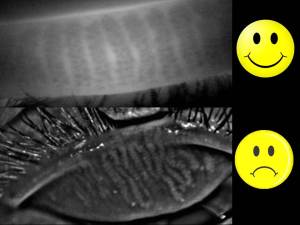
Unfortunately though, these meibomian glands are few and once blocked or inflamed can quickly become dysfunctional and die off. happysad The eyelid surface is uniquely situated close to the eyelash follicles.  Naturally occurring bacteria often accumulate in high numbers in this area due to built up dead skin, makeup and environmental debris.  This surface cannot be cleaned by commercial products with the precision required without causing harm to the eye (cleanser in the eye, removal pads can abrade the cornea, etc.).  Also the inner eyelid tissue known as the wiper is too sensitive to touch or use retail cleaning agents on without causing pain and or damage.  Basically this tissue is never really cleaned and these glands are under constant burden.

Your eyelid glands are just like your skin glands and need clinical attention to keep them functional and prevent meibomian gland dysfunction which causes up to 86% of dry eye disease patients.  In many cases it is a preventable condition, but only recently has clinical periocular hygiene emerged as a new effective maintenance treatment.  Using special dyes and instruments, the eyelid surface and glands can be treated comfortably in your eye doctor’s office.   New advances in this area allow even the most hardened blockages to be melted using prescribed heat and pressure simultaneously (LipiFlow thermal pulsation) in non-surgical painless procedure.

Bumps on your eyelid are exactly the same as pimples on your face, however the consequence of not treating it or preventing it are potentially vision threatening.  If left untreated eventual gland death can occur decreasing the stability and vitality of your tearfilm.  This puts the entire surface of the eye at risk for infection, chronic inflammation and even scarring of the conjunctiva or cornea.

Experts from around the world have now recognized meibomian gland dysfunction as being perhaps the most pervasive cause of dry eye disease around the planet.  These tiny delicate pores have a huge impact on our visual health.  Talk to your eye doctor about your glands and be proactive.  Having dedicated my career to the ocular surface and spending the last 2 years specifically developing treatments in this area, my advice is:  don’t let the symptoms start!

In good health,

Dr. Richard Maharaj OD, FAAO

Cinical Director,

eyeLABS Optometry and Center for Ocular Surface Disease

www.eyelabs.ca

twitter: @eyelabsinc

info@eyelabs.ca


Think your eyelids are clean?….REALLY! Introducing Clinical Periocular Hygiene

16 01 2014

Beauty is in the eye of the beholder. Beauty is skin deep. It’s what’s inside that counts.   These phrases gets tossed around, yet people all over the world eyelidmascaraare investing billions into the cosmetic industry now more than ever.  Men and women paint our faces and in particular our eyes to create the smokey, butterfly lashes that magazines and TV tells us we need in order to be ‘current’.   But what is happening to the tissue underneath that paint – what happens after the smoke fades away?  I can tell you from my years of clinical experience and research that the eyes aren’t very forgiving and what you’re not seeing is the decline and deterioration of one of the most important organs in your body.  Your EYES!

So after years of piling pencil liner, mascara, shadow, concealer to and around your eyes, despite your best efforts to remove all of it from the area with cleansers and patented lotions and potions, the eyelid is unfortunately ends up carrying the burden of harboring microscopic bits of make-up debris and builds a breeding ground for bacterial growth which results in an aggressive inflammatory cascade that is so sneaky that the symptoms fail to present themselves until the disease is in full swing.  That disease is LID DISEASE – it encompasses blepharitis, meibomian gland dysfunction, lid wiper epitheliopathy and more generally dry eye disease.

When is the last time you visited your dentist or dental hygienist for a teeth cleaning?  Do you brush your teeth everyday?  This concept of oral debrishygiene is well accepted and widely known that the consequences of not cleaning your teeth can lead to gingivitis and several other oral issues.  The eyelid, both upper and lower, are complex structures that have hair follicles, 2 types of differentiated skin and several glands all that are responsible for keeping your cornea nourished, clear and comfortable;  the windshield wiper of your eye so to speak.  Yet  despite the importance of this structure in keeping your vision clear and protecting the eye from infection and damage, we plaster foreign material that is in some cases toxic to this sensitive tissue and still expect it to last a lifetime?  Well what actually happens is the eye lid skin and glands (known as meibomian glands) go through a process called keratinization and atrophy.  Basically the devitalized skin crusts and grows irregularly over the gland openings as well as within the glands themselves, eventually leading to gland death in the long term.  These glands are responsible for adding the much needed oil your tears need to prevent evaporation of the tears, buffer the friction between cornea and eyelid as well as balance inflammation at the surface of the eye.  When this surface isn’t properly cleared of debris, the keratinization process is started leading to breakdown of this gentle balance that keeps our eyes comfortable and clear.  Some studies have shown that as much as 60% of adults have incomplete eyelid closure when blinking, a process that is meant to assist proper clearance of debris and epithelium from the lid surface.

damageWashing your face and eye area is important to minimize the small particles of makeup debris that can and will transfer into the eye, however the lid margin is an area that without clinical instruments is near impossible to clear with causing damage.  In fact, without specific vital dyes, similar to the ones your dentist uses to highlight plaque on your teeth, damaged tissue is actually very difficult to see even under a microscope.  Don’t be fooled when you look at your lids under a magnifying mirror and think all the make-up is gone or if you cannot see any dead skin build up.

A new innovative area in eye care is emerging to manage this preventable problem.  Dry Eye Disease is reported in scientific literature in as much as 1 in 3 adults in North America.  The problem with that number is many cases of Dry Eye Disease can be prevented by clinical periocular hygiene.  At eyeLABS Center for Ocular Surface Disease, various techniques have been developed and adapted to clean and clear this area with clinical precision and effectiveness.  A comprehensive clinical periocular cleaning is the most effective way to keep this structure healthy and hygienic to date and is a non-surgical treatment for this surface.  As clinical director at eyeLABS, our patients have benefited from  hundreds of clinical hours spent refining our non-surgical periocular techniques as well as developing new ones making our center a first of its kind facility with specialty services in periocular hygeine and treatments.  Lid margin debridement/scaling is a technique used to clear keratinized tissue after being highlighted with specific ophthalmic dyes at the eyelid surface.  Obstructed glands can be cleared which give the oils a clear path to the tear film using various expression instruments with gentle pressure being applied.  In many cases, thermal pulsation is used if the obstruction is too hardened for routine expression to clear.  This 12-minute procedure is the only FDA approved therapy for non-surgical treatment of evaporative dry eye and eyeLABS was the first optometric center in the country to acquire this technology.  Eyelash follicles and roots are known areas where bacteria and mites (Demodex) make a home and can accumulate.  This can lead to inflammation at the eyelash base which worsens the nearby tissue inflammation and damage.  Loss of eyelashes can be a sign of bacterial or mite infestation.  Up to 80% of blepharitis patients have clinically documented Demodex infestation, depite good at home hygiene.  BlephEx is a new treatment method that allows clinical removal and cleaning of this area.

Clinical periocular hygiene is a growing area of non-surgical treatments for the eyelid and periocular region which is a proven effective means of keeping the delicate skin of the inner and outer eyelid healthy and vital to preventing lid disease and maintain good comfortable ocular health. Wear and tear to this area can and does happen without makeup application as well.  Incomplete blinking, contact lens wear, environmental debris, certain medications and health problems (diabetes, thyroid, arthritis) all increase the burden to this area.  Men are just as vulnerable as women to this problem.

Like every new area of medicine, many doctors don’t even know it exists and may prescribe artifical tears, antibiotic drops, lid scrubs or other at home warm compresses and baby shampoo however none of these at home solutions are able to clinically treat this area.  Like brushing your teeth daily (or twice daily), professional grade hygiene is required for proper oral health.  The most effective therapy is a combination of good home hygiene and regular clinical periocular hygiene.  The next time you book a facial to ‘clear your pores’, consider doing your eyes a favour and booking a comprehensive clinical cleaning for your eyes.  They’ll thank you for it!

In good health,

Dr. Richard Maharaj OD, FAAO

Cinical Director,

eyeLABS Optometry and Center for Ocular Surface Disease

www.eyelabs.ca

twitter: @eyelabsinc

info@eyelabs.ca


20/20 isn’t everything: See why every child MUST have a comprehensive eye exam to prepare for school

17 10 2013

Dr. Richard Maharaj and Dr. Chris Schell demonstrate some common vision problems that children struggle with everyday in the classroom. A very important message on why comprehensive eye examinations (not a vision screening) must happen for each child prior to (or at the very least, as soon as possible) school age. Vision screenings are well-intentioned, but as shown in the video, may miss these subtle diagnoses and give parents a false sense of security. Their little eyes are starting a 12+ year marathon which will serve the basis of learning.  You wouldn’t run a real marathon without preparing your muscles and endurance would you?  Why do we assume newly developing eyes should be treated with less concern or preparation.  See a Doctor of Optometry for a comprehensive eye examination.


A Tale of Two Cities: Treating the Travelling Corneal Abrasion

12 09 2013

The kids are back in school and hopefully everyone is settling into routines like an old man into  warm bath.  With our kids back to school and hopefully learning with perfect bilaterally corrected vision (having already been comprehensively examined by an optometrist), it’s time for Eye on Eyes readers to do some learning.

The case being shared in this article is not one of an unusual pathology, but the route of management.  It speaks to the capability and compassion of optometrists across the country in helping one patient to feel comforted in a time of uncertainty.  This 62 year old female presented for the second time in 4 months with a left corneal abrasion (see photo).  Image

She had underlying epithelial basement membrane dystrophy and had been using hyperosmotic ointment at night once a week previous to this incident.  On presentation the epithelium had a crescent-shaped break consistent with her fingernail that had accidentally brushed her cornea while rubbing her eyelid.  The surrounding loose epithelium (~3mm) layed above  3+ stromal edema which created a potential for a full circumscribed abrasion with the slightest touch or blink.  She was able to keep her eye closed until coming into the clinic 15 minutes after the incident.

Certainly a worrisome cornea with the potential for infection to set in, however there was no evidence of contamination of the wound and there hadn’t been a lot of time for the eye’s natural flora to cause further insult.  Managing this required wound protection and prophylaxis measures to prevent infection.  The monkey wrench was that this patient was flying to Calgary later the same day and I was left with a potential ulcer, scarring and related vision loss if this wasn’t followed promptly and compliance with my treatment wasn’t followed.

This scenario required some “outside of the box” thinking and in fact outside of the province thinking.  Luckily, my esteemed colleague, classmate and friend Dr. Dwayne Lonsdale who practices near Calgary (North Hill Optometry) was just a facebook message away and was available to follow up and be her on-call travel optometrist while she was in his area.   With the patient’s consent, I sent Dr. Lonsdale (http://www.northhilloptometry.com/) the above image (taken using my smartphone behind slitlamp) for reference and follow up until she could return to my care back in Toronto.

Once her travel-care was arranged, I placed a bandage contact lens on eye, provided her with  antibiotic topical coverage and Muro 128 qid + ung qhs and sent her safely into the slit lamp of another.  With recurrent corneal abrasions it is important to heal the wound first by protecting it from chronic insult.  In this case repeated mechanical trauma of blinking would cause this epithelium to slide right off and leave an open wound waiting for a biological enemy to invade and infect.  During this time treat with topical antibiotic coverage (4th generation qid) and hyperosmotic agent to reduce edema.

Dr. Lonsdale reported her progress and removed BCL by day 3 and wound recovery was excellent.  BCVA had improved from her initial 20/40- to near 20/20.  Once the wound had closed, a topical antibiotic/steroid was added to reduce inflammation further while retaining coverage.Image

On returning from her trip to see me, her prescribed medications were reduced to hyperosmotic ointment nightly and non-preserved 1% hyaluronic acid to replenish the epithelium.  She is fully recovered and eternally grateful for the care she received at home and while travelling in Canada.  We are discussing options to prevent further RCE by using oral doxycycline combined with hyperosmotic ointment nightly to reduce ocular surface inflammation.

What is interesting here is that without smartphone anterior segment photography, social network communication and the close optometric community that we have, I would not have been comfortable with this patient travelling and would have cautioned her to postpone this trip.  Leaving a BCL on an eye with an open wound with the potential for an opportunist infection and sending her on a plane without confirming receptive eye care on her arrival would be a liability to say the least.  But instead she travelled confidently, she healed and we all learned what is possible when people work together.

Here’s hoping our kids will learn to do the same this year!

In good health,

Dr. Richard Maharaj OD, FAAO

Director of Optometry,

eyeLABS Inc.

www.eyelabs.ca

twitter: @eyelabsinc

rmaharaj@eyelabs.ca


Dr. Chris Schell discusses Ontario’s Eye See Eye Learn Program on CTV news

28 08 2013

Dr. Chris Schell is featured on CTV Barrie discussing the Eye See Eye Learn program in Ontario. He discusses the importance of children’s eye examinations for school aged children. Dr. Schell has a special interest in Vision Therapy and Pediatric Optometry.


A Child’s Vision: How important is it to you

19 08 2013

Only 14% of children under 6 in Canada have had a comprehensive eye exam yet 80% of learning is visual. In preparation for a 12+ cycle of education is this how do we equip our kids? We should expect more. Many common learning disabilities have been shown to be hidden undiagnosed visual dysfunction. Schedule your child’s eye exam today!


Counterknowledge: Is Dry Eye a Disease or a Syndrome?

5 08 2013

The answer:  IT IS A DISEASE!

Language is important and how we treat a medical ailment depends very much on what we call it.  Terminologies like disorders, syndromes and diseases get mixed up and misused and interchanged depending on the literature or even the medical professional you are speaking to.   Defining a condition correctly will change the attitude of the patient suffering from it and the doctor treating it.  Brampton-20130205-00310Dry eye is one such disease that has been misrepresented as a syndrome in many arenas but let’s take a look at the definition of a disease versus a syndrome.

Syndrome:  a collection of signs and symptoms known to frequently appear together but without a known cause.  This grouping generally characterizes a disease or disease process

Disease:  a morbid entity characterized usually by at least two of these criteria:

  1. Recognized etiologic agent (cause)
  2. Identifiable group of signs and symptoms
  3. Consistent anatomic alterations

Dry eye disease, also known as Keratoconjunctivitis Sicca, is the term used by the internationally recognized Tear Film and Ocular Surface Society (TFOS).  It has very clear and identifiable signs and symptoms, anatomical changes are both diagnostic and prognostic of the disease itself.  The cause, or etiology, of dry eye is an often debated subject but as a culmination of decades of scientific study, it is well agreed that it can be distilled into one or a combination of aqueous deficiency, lipid or oil deficiency and/or cicatricial (scarring).  It is also generally accepted that dry eye is an inflammatory disease, which is why the majority of pipeline drugs are targeting inhibition of specific inflammatory pathways.

Why is this conversation relevant?  Too often a ‘syndrome’ get’s swept under the rug or trivialized by medicine and pop culture.  We are swift to group symptoms together and call it a syndrome which may be reason enough to take this side-stepping approach.  However when a real and clearly defined condition affects over 25 million US adults and over 100 million people world wide, AND science has elicited cause and effect then it should become an imperative to give it ‘disease’ status; not to scare or induce fear, but to appropriately identify and manage the process.

The next time you meet someone that has dry eye disease (DED), don’t define that person by the disease but rather understand the impact that it has had on her/his life.  Ask them how many doctor’s have actually given it the attention it deserves.

In a survey of 100 patients at eyeLABS Center for Ocular Surface Diseases, the average number of eye physicians/doctors the patient had consulted for DED was 3 prior to seeing me.  I intend to be their last.

sidenoteSideNote: The Ocular Surface is Skin – Treat it that way

Dry Eye Disease is a skin condition, not unlike many dermatological conditions.  The lid surface, meibomian glands and corneal tissue are variations of epithelium and sebaceous glands which will age, like the dermis does.  The lengths of cosmetics, creams, lotions and potions for the skin can help to preserve our skin, but what about the eye?  The science at eyeLABS is founded in ocular surface skin preservation and sustenance.  Lid Margin Debridement (click here for related article) and clinical gland expression provides a basis for the spectrum of treatment options and maintenance procedures available at our clinic.  Contact lens wearers in particular should actively seek these types of treatments out as they are more likely to develop lid related inflammatory conditions (lid wiper epitheliopathy) that directly impact the glands, cornea and therefore dry eye disease progression.

Dr. Richard Maharaj OD, FAAO

Director of Optometry,

eyeLABS Inc.

www.eyelabs.ca

twitter: @eyelabsinc

rmaharaj@eyelabs.ca


Be Proactive on Reducing Wrinkles – add comfort to your eyes!

25 07 2013

“Epidermal atrophy and structural changes observed in the Dermal-Epidermal Junction with aging may be, by some extent, related to daily and repetitive skin deformations all along the life span”

Skin Res Technol. 2013 Jun 25

Translation:  Wrinkles and the aging skin are related to constant stretching and relaxing of the skin.  This may sound like common sense, yet patients everywhere still engage in life behaviours that are known to hasten wrinkle formation.   Smoking, tanning, high sugar diets all are big contributors to the aging skin but while changing your habits may take some work, why not eliminate some factors that are out of your everyday control.

crowsfeet

Squinting your eyes – a very common ocular reflex to external factors such as bright light, blurry vision and ocular discomfort.  Because it’s a reflex it is not considered a bad habit, but rather a mechanism your body employs to protect the ocular surface and or enhance vision when stimulated by the above factors.  Here are some very easy steps to reduce your stimuli to squint:

  1. Wear corrective eye wear when needed – this is by far the easiest step to take to avoid squinting.  It has the added benefit of clearing your visual world.
  2. Wear your sunglasses – UV exposure worsens this cause of squint because it can accelerate the aging skin.  If you’ve ever lost your sunglasses and spent even a lunch hour without them, you’ll likely have a headache all afternoon.
  3. Treat your ocular surface keeping it wet and comfortable – Dry Eye Disease (DED) is a leading cause of eye discomfort.  When left untreated the natural course of this disease will worsen leading to further eye muscle contraction.

The treatment options for dry eye have changed dramatically as experts world wide agree that meibomian (oil) gland dysfunction (MGD) may be the leading cause of dry eye in the world (TFOS 2011).  Knowing the mechanism behind this condition allows it’s treatment to be more targeted and therefore more effective.  Each of the four eyelids are home to the 20 to 40 oil glands and optimal functioning is achieved when the glands are absent of any obstruction and the lid surface is properly cleared of debris and devitalized epithelium.  The eyelid margin (the area behind your eyelashes towards the eye) is the only area of our bodies that doesn’t get cleaned regularly or thoroughly and because of its proximity to the cornea is a dangerous place to try to clean at home with q-tips, brushes or wipes.  Corneal scratches can become infected and are extremely painful.  Also, over the counter cleansers are still insufficient to removed devitalized skin and debris which can only be seen under microscopic examination with ophthalmic vital dye staining.LOM_mgo

eyeLABS Center for Ocular Surface Disease offers an array of services to treat and maintain your glands and lids for optimal functioning:

  • Lid Margin Debridement – and exfoliative process for the eyelid surface
  • BlephEx – a microdermal exfoliation of the eyelid surfaces and eyelash roots
  • LipiFlow Theramal Pulsation – the only FDA approved treatment for MGD.
  • Topical and Oral Medicines – although we focus on enhancing your body’s natural functioning there are cases that require specific medicine to treat underlying inflammation and/or infection

In this way, we can limit your stimulus to squint and help you widen those eyes and lose the wrinkles!  Instead of spending countless dollars on Botox and fillers, be proactive and prevent unnecessary “epidermal atrophy,” also known as wrinkles!

Dr. Richard Maharaj OD, FAAO

Director of Optometry,

eyeLABS Center for Ocular Surface Disease

www.eyelabs.ca

twitter: @eyelabsinc

rmaharaj@eyelabs.ca