Taking the Fear out of treating Herpetic Keratitis

6 03 2013

Fear.   The word itself causes one to take pause and hold back.  For doctors, fear of litigation likely tops the list of reasons to refer patients out but fear of making a disease worse  has historically allowed half truths about treating anterior eye disease to thrive.  For instance the fear behind using a topical steroid by both family medicine and even ophthalmology and optometry has hindered its use to this day – even when it is indicated for treatment.  This article will discuss a specific ‘sore’ spot in the eye bank, HSV and HZO, and the role of topical steroid administration in quelling this persistent nemesis.

HSV is carried by 90% of people, however most remain dormant and don’t show up in our exam chairs.  When they do however, red flags and alarm bells immediately start to ring because the ability of this bug to thrive has caused corneal scarring  and blinded many since its first human to human identification by Vidal in 1893.  Although patients assume sexual transmission when they hear “herpes,”  eye related infections are often not primary and therefore not sexually transmitted at all.  The stigma, like the virus, has survived despite our best efforts.

The inflammatory stage of herpes simplex keratitis occurs because of the coating of the virus itself produced from this clever virus’ protein coding.  One protein in particular similar to human corneal protein is called UL8 which triggers an immune response.  Because UL8 is similar the normal corneal proteins, the immune system begins to attack healthy tissue in addition to the virus itself, triggering CD4+  T cells which release chemokines and Th1 cytokines resulting in a destructive inflammatory cascade. Other supposed sources of inflammation are CD8+ T cells which are more destructive and lead the corneal scarring that has come to be associated with HSV end stage.

Epithelial Involvement:

HSVk

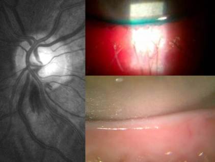
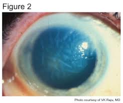
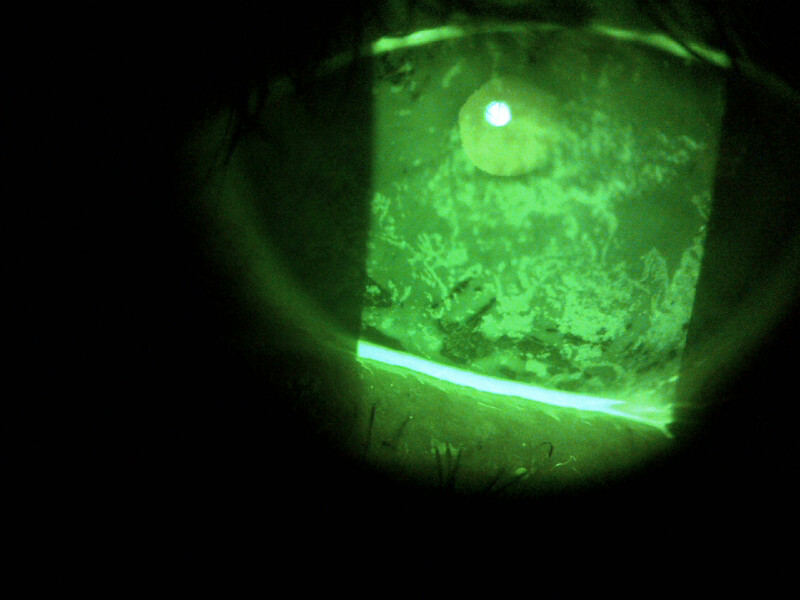
Differentiating is key.  Epithelial involvement can be small or large lesions with that typical dendritic appearence, but without a terminal bulb glowing with an additional vital dye (Rose or Lissamine), it can be easily misdiagnosed.  If you don’t have an additional dye and are convinced of bulbs then back up your diagnosis with corneal sensitivity testing.  Dental floss – the old nylon type – or even a cotton tip that has been teased out can serve as an vital tool at confirming your diagnosis.  Test the good eye first and then the affected eye.  The aesthesia score will be markedly reduced and this is a positive diagnosis of HSK.  Clinical history will generally agree with these 2 test results.

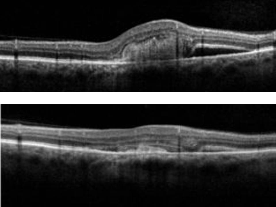
Interstitial Keratitis vs. Endotheliitis:

Stromal or interstitial keratitis (immune mediated) will be just that – limited to the stroma and will NOT involve the epithelium or the endothelium.  A thin beam on your slit lamp will help to isolate the haze and only in this isolation can you diagnose as interstitial.  Often misdiagnosed as interstitial keratitis is the disciform lesion.  A true disciform lesion (without epithelial invovement) will ALWAYS involve the endothelium and is in fact an endotheliitis.  HSV endotheliitis comes in 2 other forms diffuse, and linear, based on the pattern of endothelial dysfunction and stromal edema.  Typically this is accompanied by an anterior chamber reaction and elevated IOP second to trabeculitis.

HSVendo

Of course, with epithelial involvement the use of a steroid is contraindicated however doctors should not fear or hesitate its use in cases with intact or non-necrotizing epithelium.  Interstitial HSK and endotheliitis require a topical corticosteroid administered in addition to oral antivirals to suppress the immune response.  Now that said, we’ve all been burned by a case in the past so what will happen if a steroid is applied to an epithelial case?  This is where your clinical judgement and direction is key.  Follow up in 1-3 days and clearly instruct this patient that should vision or symptoms change for the worse at all, to call the clinic immediately or head to urgent eye care if after hours.  An incorrect use of steroid on a small missed lesion will not cause the eye to explode but will serve as a provocative test.  Yes the lesion will expand and the virus will accelerate shedding.   I’m not condoning or advising the use of steroid to diagnose HSV, however I will follow that by saying it is a manageable ship to turn around with topical antivirals, (zirgan is very effective) provided it gets caught early and managed aggressively.

Topical Treatment:

HSV keratitis:

  • Viroptic (trifluiridine) q2H x 1 wk or until epithelium is healed and thereafter qid x 1 wk.
  • Zirgan (gangcyclovir)5x daily x 1wk or until epithlium is healed and thereafter tid x 1 wk

Zirgan is an excellent new additional to the arsenal and friendly to the ocular surface.

Oral Treatment:

  • Zovirax (acyclovir) 400mg five times daily for seven to 10 days
  • Valtrex (valacylovir) 500mg three times daily for seven to 10 days
  • Famvir (famciclovir) 250mg three times daily for seven to 10 days.

HZO keratitis:

  • Zovirax (acyclovir, GlaxoSmithKline) 800mg five times daily for seven to 10 days.
  • Valtrex (valacylovir, GlaxoSmithKline) 1,000mg three times daily for seven to 10 days
  • Famvir (famciclovir, Novartis) 500mg three times daily for seven to 10 days.

Interstital or Endotheliitis:

Add topical steroid (lotemax or prednisolone have demonstrated equivalent efficacy) q1-2h and taper appropriately as resolved which can be for weeks or months.  This is extremely key in reducing the immune response.  Without topical steroid it can persist and prolong viral shedding and even increase the risk of necrotizing and or scarring.

Now this certainly isn’t an A-Z coverage on HSV or HZO  – geographic and neurotrophic ulcers are both noteable subsets which generally occur in chronically untreated or misdiagnosed cases and have extreme visual consequences.  I’ve chosen to reserve that discussion for another thread.  Managing the ocular surface is always paramount so Viroptic preserved with thimerasol is certainly a thorn, which is why Zirgan offers a better and more effective alternative.   Of course lubricate with preservative free drops during this period and be careful to instruct about drop insertion.  If the patient is doing what you’ve instructed, the only reason the keratitis will persist is because they are poking themselves with the dropper tip and causing their own keratitis!

Dr. Richard Maharaj OD, FAAO

Director of Optometry,

eyeLABS Inc.

www.eyelabs.ca

twitter: @eyelabsinc

rmaharaj@eyelabs.ca


Counterknowledge: “I only wear my contacts ‘once in a while’ so I usually replace them every 2 to 3 months”

20 02 2013

Do you drink milk past the expiration date?

Not likely, and if you do you’re probably not happy about it.  Contact lenses are tiny thin pieces of hydrated plastic that, like every other material in the world, are not impervious to getting dirty.  Despite pharmaceutical companies’ best efforts to create the perfect cleaning systems, even the best cleaning agent (hydrogen peroxide based systems) for both contact lens and ocular surface still doesn’t clean everything.  Protein sticks to the contact lens surface and over a period of days to weeks will denature or break down.  This denatured protein does not agree with the ocular surfaces  and can cause a host of complications that may not have immediate symptoms or signs.

17 million people in the US alone have Contact Lens Induce Dry Eye or CLIDE (Ramamoorthy P, 2008).  The peak age of contact lens wearers is in the mid-20’s and Brampton-20130205-00310

How often do you change your oil in your car?  How often do you and your eye doctor discuss  your eyes oil and how it relates to contact lens comfort and every day optical optimization?  Japan is home to the world’s highest number of LASIK surgeries performed at Shinigawa LASIK centers with over 1 million surgeries to date.  In order to optimize visual and surgical clarity each patient undergoes LipiFlow thermal pulsation, a new standard for treating ocular surface disease – specifically dry eye.  In December 2012, Shinigawa set a new standard in LASIK by treating 1000 patients with the LipiFlow system.   This precedent shouldn’t be isolated to those paying for premium refractive surgery.  The amount of money you invest in contact lenses over a lifetime is likely more then you would pay for lasik, so why does the ocular surface not matter as much?

Proper maintenance and therapies for you eyelids, cornea and contact lenses are crucial for comfortable clear vision but the wide and sometimes careless availability of contacts through online stores and big box environments have turned contacts into a commodity and with that comes this pitfall – ‘If I can shop around for the lowest price like a pack of gum then it must not be worth taking care of.  Right?’  But are your eyes a commodity that is as easily replaceable?  If you knew 10 years from now that you wouldn’t be able to tolerate your contact lenses would you do something different today?

Here’s what you can do:

1) Wear single use lenses otherwise known as daily disposables   Get rid of your case and solution and use it once and then toss it.  The healthiest contact lens is no lens at all.  The second healthiest is a daily disposable – sterile fresh lenses in the eye every day.

2) Treat your eyelids well – if you wear make-up, smoke, work in an office or use a computer for more then 4 hours a day then statistically your eyelids and blink are likely to be contributing to lid disease down the road.  Talk to your doctor about ocular surface health and options for maintaining it.  How often do you get a facial?   Your eyelids deserve the same!

3) Don’t shop downwards.  Consider your contact lenses and vision choices the way you would consider LASIK – you would make your decisions based on risk, doctor experience, equipment safety and statistical likelihoods first before looking at price.  Just because you can find them cheap online, does not mean they were created equal.

sidenote

SideNote: LipiFlow is available in Canada

LipiFlow is the only FDA approved in-office procedure that has demonstrated clinical effectiveness in treating the ocular surface, specifically meibomian gland dysfunction (MGD).  Although offerred to pre-operative patients going through lasik and cataract surgery in large refractive surgery centers, eyeLABS dry eye clinic is dedicated to the ocular surface  and is located in Brampton, Ontario.  A 12-minute non-surgical procedure has provided patients with relief of symptoms for up to 15 months as reported in clinical trials.  Making this available to everyone will make a difference to contact lens wearers and non wearers alike – Dr. Maharaj is the first optometric clinic in North America to acquire this technology.  Call 905-456-9333 to discuss your dry eye options.

Dr. Richard Maharaj OD, FAAO

Director of Optometry,

eyeLABS Inc.

www.eyelabs.ca

twitter: @eyelabsinc

rmaharaj@eyelabs.ca


Revitalizing the Eye Waterfront: Treating Lid Disease

4 02 2013

Managing patients with dry eye disease (DED) over this past year has transformed the foundation for my clinical decisions with regard to the ocular surface.  Going back to basics has  had a profound impact on my patients with ocular surface disease (OSD).  In November 2012, I wrote a piece on eyelid plaque and its role in OSD which spread to eye physicians globally, all asking about how and why this hasn’t been done before and if I have any examples that I could share.

The answer as to why Line of Marx (LOM) debridement technique isn’t well documented lies in the fact that LOM isn’t robustly described in scientific literature nor has significant emphasis been put on this area in academic institutions or research arenas.  Well if it is not trickling down to clinicians yet, it soon will.  Already, the 2012 American Academy of Optometry meeting in Phoenix featured a lecture being leading experts Dr. Kelly Nicols and Dr. Caroline Blackie in which Dr. Blackie mentioned LOM debridement as an effective adjunct for MGD patients.  In a recent article in the Review of Optometry, a Lifetime of Dry Eye, Dr. Cheryl Murphy mentions this technique while quoting Dr. Blackie.  I would keep your eyes and ears open at CE and research meetings in 2013 for works related to LOM as a source of chronic  DED.

before_afterLOMdebride

In the mean time I will share an example of a recent case of a longstanding chronic DED patient.  This female patient in her late 30’s underwent LipiFlow Thermal Pulsation 7 months ago and has been relatively asymptomatic since that time.   She uses oil emulsified drops once daily.  Prior to her therapy, she presented with mg atrophy and notching L>R, but symptoms were always greater on her left side which happened to be the side with more mg notching and atrophy.  She has poor lid apposition (lid seal) and is a partial blinker – which is exacerbated at night by exposure.  On Friday’s presentation she reported significant burning OS of gradual onset throughout the week.  Clinically she presented with moderate to severe superficial nasal and temporal keratitis.  Her LOM was anteriorly displaced (no progression since LipiFlow) and had moderate devitalized epithelial accumulation.  I performed LOM debridement and meibomian gland expression to her left eye and advised to increase lubrication to twice a day (vs. once daily).  Historically she responded well to this, however if this was an initial presentation on a new patient, I would add topical antibiotic for coverage and add an overnight ointment.

The patient returned 3 days later for follow-up and as can be seen in her slit lamp image with fluorescein staining, the keratitis was vastly improved and symptoms were resolved.  And so it goes with many of my dry eye patients.   A burdened cornea needs less burden which is why I did not add copious lubrication, and therefore copious preservatives, to the regimen.  This technique offers a management alternative to loading the ocular surface with agents that may mask the true etiology of the problem.  Ideally this patient should do well with a preservative-free option, however she hasn’t found relief in this in the past and was using PF drops 10-15 times a day with limited relief.  With lid therapy we managed to lighten the financial burden as well

This is a chronic patient, and by no means am I or is she expecting a cure-all of her OSD.   The ‘after’ is still not perfect, but it is a safer cornea.   We will continue to work together to as doctor and patient to customize her therapy all while maintaining her lid surface regularly.

LOM_mgoWhen I originally read Bron’s work (Ocul Surface, April 2011) about  the solute gradient created in hyperosmolar tears and how it relates to LOM and MG damage, it was hypothetical at best to me and I had a hard time picturing it.  In this picture, the process comes to life as you can visualize the LOM with little ‘teeth’ straying periodically toward individual MG orifices.  This is the path to damage and helps to explain why a notched lid has that v-shaped appearance – it is not unlike a high speed river creating a pathway clearing anything in its way to get to the drain, which in this case is the meibomian gland.  Once this pathway is created, the gland is now at the mercy of inflammatory factors and the solute gradient created at the base of the tear film.

Clearing this devitalized epithelium from this pathway may serve to disrupt the cycle and slow the progression of lid disease.  With research and new drugs being aimed at reducing ocular surface inflammatory mediators, eye care providers can make a difference by monitoring this tissue closely and revitalizing it periodically.

sidenote

SideNote:  All the clinical images were taken with a smartphone through a slit lamp.  This is a simple yet effective way to monitor the anterior segment and can be done with relative ease.  There are a number of smart phone adapters available, however none that are universal.  The variations in eye piece diameter make this difficult, however I managed to align these images manually.  


Dr. Richard Maharaj OD, FAAO

Director of Optometry,

eyeLABS Inc.

www.eyelabs.ca

twitter: @eyelabsinc

rmaharaj@eyelabs.ca


Counterknowledge: Will wearing my glasses make my eyes worse?

29 01 2013

The answer:  NO!

Countless patients, particularly parents of my pediatric patients, report reduced wearing time or frequency of spectacle wear in order to prevent their eyes from weakening.  Somehow, the urban legend that ‘wearing glasses too much will make my eyes worse’ has perpetuated through the hallows of time and has blossomed into a weed that I and many colleagues are constantly trying to eradicate through education.

Let’s look at an analogy – Does wearing your shoes 2 sizes too big make your feet bigger?  Does not wearing shoes make it easier for you to walk?  Does making it easier for you to walk make your legs weaker?  These all seem like ridiculous questions, but it is this rationale that people rely on when thinking about wearing eye correction.  It just isn’t true or supported by scientific evidence.

Now there is some evidence that a low level of uncorrected nearsightedness (myopia) may be beneficial for close work and not using eye correction for these instances may have a positive impact on myopic progression, however by and large this theory is still being tested.

Some people who have binocular vision development issues require more intensive direction as to when and when NOT to wear glasses.  In these cases your eye doctor will communicate this specifically during your examination.

When parents unilaterally make the choice to reduce the wearing schedule for their kids based on this urban myth,the effects on visual development and subsequent academic performance can be significant.  80% of our learning comes from visual learning and not having optimal vision has far reaching, and a well documented negative impact on educational development.  Often I hear “my child’s eyes are lazy so I just want them to work harder.”  My advice to anyone that has said this or been told this is to really think about this concept.  More importantly, as eye doctors our goal is to use evidence based approaches in tandem with our extensive knowledge of the visual system to make a judgement on how to treat your eyes and vision.  The recommendations of your eye doctor are not made on a whim and are prescribed for your specific visual system based on your exam findings.

Bottom line – Wear them and wear them often.  Give your brain the vision it craves.  The clearer an image is focused on the retina, the better the resolution of the visual stimulus to the brain.  This means that the neural network developing between the photoreceptors in the eye to the optic nerve to your brain is more intricate and allows a better potential for vision.  Some people end up feeling their uncorrected vision becomes worse after wearing glasses for some time.  What is actually happening is your brain is getting used to 20/20 and is preferring it.

  • A quick test:  To know if your vision is truly getting worse try this.  Put your glasses on and stand 6 kitchen tiles away from your digital clock on the microwave or stove.  Cover each eye and read the time.  Use this as your baseline and compare it monthly in exactly the same manner.  If the clock becomes blurry WITH GLASSES ON, then your vision has changed.

To play devil’s advocate I understand that it is conceivable that because your doctor dispenses glasses that you may consider his or her recommendation to wear them self-serving.  If you feel this is the case, then seek out a second opinion and validate (or rule out) your concern.  Steering yours or your child’s visual management based on misinformation is dangerous, no matter how many times you read it on the internet or have friends that ‘have the same eye problem.’  No two eyes are the same – not even your own!

Here are some points to clarify with your doctor when being prescribed glasses:

  • Frequency and Duration: When and for how long should I wear them?
  • Alternatives:  What other vision correction options do I have?  Contact lenses?
  • Adaptation:  What are normal symptoms should I expect when wearing my glasses for the first time?  What is considered abnormal?

sidenoteSideNote: Online shopping

The next time you consider purchasing your eye wear online, consider this:  Who is responsible if something goes wrong?  What value do you place on the service that comes along with your eye wear.  For some, this point may be moot however anyone that has had trouble adapting to the vision of a new pair of glasses or a new snug frame will tell you the value of having your optometrist or optician provide the ophthalmic service is worth it.  If you’re willing to give up service then consider a September 2011 study by a research professor at Pacific University College of Optometry in Oregon that found that 44.8% of eyewear ordered online FAILED at least one parameter of optical or impact testing (Click here to see study).

Dr. Richard Maharaj OD, FAAO

Director of Optometry,

eyeLABS Inc.

www.eyelabs.ca

twitter: @eyelabsinc

rmaharaj@eyelabs.ca


Lessons in eye care

24 01 2013

As the content of our textbooks become outdated keeping up with current opinions in eye care can be daunting.  Combined with that is a complacency that some doctors have that they will somehow stay current through osmosis.  The reality is that keeping abreast of all the general and sub-specialties within optometry and ophthalmology is near impossible so when I stumble upon a GREAT resource, the educator in me must share it.  Dr. Larry Alexander OD FAAO, a great friend and a pillar in the optometric community has started a wonderful website eyelessons.com.  I encourage all of my colleagues in health care to register and use this sponsor-free resource.  Like Eye on Eyes, this is a living breathing community that evolves as current eye management evolves.  Click here to visit Eye Lessons with Dr. Larry Alexander, author of Primary Care of the Posterior Segment (among many others).


Eye on Eyes Top 3 Eye Supplements for 2013, By Dr. Maharaj

15 01 2013

The nutriceutical industry is bombarded with ‘miracle medicines’ claiming to cure all that ails you, however without any regulating body these over-the-counter non-prescription pills are too varied for the consumer to make sense of when faced with 5 feet of choices at your local superstore or pharmacy.  Now, the reality is that some molecules actually do have merit and have been shown in repeated studies and world-renowned institutions like John Hopkins Hospital to have medical efficacy in a variety of conditions.  In this article, I will reveal my top 3 picks for nutriceutical supplements for the eyes for 2013 and why. These picks are based on my research and my clinical work with these supplements.  A little background here:  I have worked with A LOT of supplements over my career and I have  scrutinized claims made in scientific literature in clinical settings. I chose them after a detailed due diligence on my part and to be clear I have no financial interests or ties to any of the listed products.

Omega 3 (PRN – Physician Recommended Nutriceutical) : Dry Eye Disease, AMD

Now, I’m being very specific here about the PRN formulation for a few reasons which will be explained shortly.  Omega 3s have been historically shown to be essential nutrients derived from dietary consumption.  Our bodies cannot produce or store this molecule so where we get it is the only variable we can control.  Natural sources of O3’s include plants and animals each with their own unique form.  Plant sources, flaxseed being most recognized,  contain alpha-linoleic acid and marine animals contain the triglyceride form.  From a physiological perspective alpha-linoleic acid (ALA)  forms are less bioavailable then triglyceride (TG) forms because the enzyme required to convert ALA to O3, delta-6 desaturase, can be pre-occupied with Omega 6 conversion of pro-inflammatory factors.  That’s right –  flaxseed in the average omega 6 rich North American diet will not convert to EPA/DHA as readily and will likely add to pro-inflammation.

Triglyceride O3 are available as unpurified, ethyl ester (EE), and re-esterified TG (rTG) forms and with each step the cost of production rises – For good reason.  Unpurified forms, the least expensive, are just that and have all the toxins and impurities that come with pressed oil directly from fish.  EE forms, which  cost slightly more, have been pressed and heat purified using ethanol to convert the TG to and EE which is less bioavailable (Dyeberg J, Nutrition 1995).  rTG forms go through the same process as EE but are the ethanol is removed (re-esterification) and replace with the triglyceride backbone.  This critical extra step does add to the production cost, but the absorption of  rTG O3 nears 100%.  It is this small difference that minimizes the gastrointestinal distress, heartburn and fish-burps you experience with many over the counter EE forms.

Dry Eye Disease (DED), macular degeneration (AMD) and other ocular diseases with known inflammatory roots benefit from this supplementation.  A recent study by Dr. Gregory Smith MD demonstrated that 82% of patients treated with rTG (1,680 mg EPA/560mg DHA/1,000mg vitamin D) had changes in their oil gland secretions at 8 weeks.  rTG O3 is the closest to direct pure fish derived O3 and is a key differentiator in quality of the vast array of choices.  For my ocular surface patients/dry eye patients, I see PRN formulation of rTG O3 being a standard all other formulations should be measured against at a minimum dosing of 2000 mg/day.  Patients should consult there eye physician prior to taking particularly those on blood thinners.

 Resveratrol (Longevinex): Wet/Dry AMD, Cardiovascular Tendencies

Once touted as the next fountain of youth due to myriads of publications demonstrating its cardiac and ‘anti-aging’  health benefits received a big blow to its momentum in January 2012 when Dr. Dipak Das was accused of scientific fraud in a dozens of his publications asserting its efficacy.  A midst this brewing controversy, Dr. Stuart Richer OD PhD FAAO has been studying the molecular effects of this drug on the aging eye – specifically eyes with age-related macular degeneration.

Resveratrol is a molecule extracted from the skin of grapes in red wine. Only 1mg trans resveratrol in 5 ounces of best red wine and therapeutic doses require 100mg so before and afterdon’t think you can drink this into your system.  RV modulates protective pathways against oxidative stress,

excitotoxicity, inflammation, DNA  damage, and apoptosis or cell death (Richer, S).    The mechanism of action is multifaceted, however recently facilitation of survival of endogenous stem-cell by this small molecule was achieved by breaching the protective retina barrier.  Stem cells can be implanted, however undifferentiated stem cells are available in adult tissue when it becomes damaged.  RV may hold a key in enhancing this process.  It also has been shown to down regulatethe mRNA controlling  vascular e

ndothelial growth factor gene (VEGF) which is a welcomed alternative to intraocular injections of anti-VEGF drugs for wet AMD patients  (Richer S, William S, et al University of Illinois (UIC), Retina Service, Eye and Ear Infirmary, Chicago, IL 2011).  The image to the right is a before and 18 days after resveratrol administration (no other forms of treatment).  Normally this reduction in lesion size can only be achieved through intraocular injection of anti-VEGF.  I do however acknowledge although the natural course of wet AMD tends to worsen, there are cases of self resolution without treatment.  In Dr. Richer’s study, however, he demonstrated the repeatability of this finding in 18 patients.

As a practitioner, the potential of this molecule in specific vascular diseases like AMD is immense.

Patients undergoing medical management of AMD should be aware of the brand Longevinex because of its micronized/microencapsulated powder form which allows for slow release of the trans RV.  This drug specifically has been used in several eye related studies and the inclusion of vitamin D3 is a key component.  I would argue that we have yet to see the full ability of this molecule to improve vascular health.

Macular Carotenoids – Lutein(L), Zeaxanthin (Z) and meso-Zeaxanthin (MZ) (MacuHealth): Dry AMD/pre-Dry 

The yellow pigment found at the macula is a distribution of carotenoids L, Z and MZ which are 3 of over 700 found in nature.  Together these 3 pigments are known as the macular pigment (MP).  MP acts as a filter for blue light and as an antioxidant which is believed to protect against AMD (Snoderly, 1984).  In fact studies have indicated that the combined ability of the 3 MP’s to quench singlet oxygen/free radicals therefore reducing oxidative stress is greater than the sum of their parts (Binxing Li, et al 2010).

Several studies have examined serum concentration of MP’s in response to supplementation and demonstrated that the combined consumption can very depending on the cocktail in question.  The British Journal of Nutrition in September 2012 published a paper demonstrated a specific proportion (10 mg L,2 mg Z and 10 mg MZ) produced the highest serum concentration in a small group of patients using HPLC (high performance liquid chromatography).  By optimizing serum levels of MP, this makes it more bioavailable for capture by the retinal tissue.

MP density at the macula has been shown to be diminished in patients with AMD and it is possible that patients with a reduced MP profile may be at increased risk for macular disease.  The AREDS II study, yet to be published, has included L and Z as treatment arms and preliminary results confirm the importance of carotenoid supplementation.  Unfortunately MZ wasn’t included in AREDS II, however with the growing body of science supporting the use of all 3 carotenoids vs. L and Z alone it stands to reason to supplement all three inclusively.  MacuHealth is a unique supplement that combines all MP’s which is key as MZ is hard to come by in supplement form.  As the study of MP continues to evolve, MacuHealth seems to remain at the forefront of preventative treatments for macular disease.

And that rounds out my top 3 nutriceuticals to watch and perhaps consume in 2013.  My approach to health care is evidence-based and to my patients and colleagues alike, I do put a great deal of work into what I recommend to you.  I encourage you to seek alternatives but to weigh them out in addition to determine if it is truly an effective alternative.    As always do consult your eye care physician when deciding whether supplementation is warranted and/or safe.

Dr. Richard Maharaj OD, FAAO

Director of Optometry,

eyeLABS Inc.

http://www.eyelabs.ca

twitter: @eyelabsinc

rmaharaj@eyelabs.ca


Necessary diabetic eye exams skipped despite funding

8 01 2013

Necessary diabetic eye exams skipped despite funding

Delisting routine eye examinations for adults had the unintended consequence of reducing publicly funded retinopathy screening for people with diabetes, researchers in Ontario say.

Fewer people with diabetes are getting eye exams to prevent complications, which researchers call an unintended consequence of the Ontario government’s decision to delist the exams for other adults.


Regenerative Medicine with Corneal Transplants

8 01 2013

Regenerative Medicine with Corneal Transplants

Ocular Systems, Inc. (OSI), Wake Forest Baptist Medical Center and the North Carolina Eye Bank have formed a new company based on a promising new technology aimed at engineering replacement corneas in the lab for transplantation.


Click here to read full article


Putting Pressure on the Eyelid: Glaucomatous Lid Disease

4 01 2013

Chronic Disease – A burden that many of our patients carry.  Curing a condition isn’t nearly as common as managing it however eye physicians shouldn’t lose sight of the bigger picture.    Treating glaucoma, a common ocular chronic disease, has evolved into more then IOP measurement with a Schiotz tonometer and measuring cup/disc ratios with a direct ophthalmoscope.  We monitor visual field progression, RNFL loss, structure function maps, and even multifocal-ERG may hold a promise in managing this neuropathy.  All this even though the only factor we are able to modify is IOP.  Interestingly, in the race to reduce the medicinal load on our patients, we have inadvertently been accelerating damage to the lid surface – most specifically the line of Marx (LOM), meibomian gland orifice (MGO) and acini (MGA).

A recent study at  University G d’Annunzio of Chieti-Pescara, in  Chieti, Italy, in vivo laser scanning confocal microscopy was used to reveal morphological changes to the MG in patients being treated for glaucoma compared to a control group.  Specifically the eyelid margin epithelial cell density, mean acinar density (MAD) and area (MAA), glandular orifice area, secretion reflectivity and non-homogeneous appearance of interstice and acinar wall were observed in these groups.

Patients that were on 2 or more drugs had the greatest morphological changes to the lid surface, specifically preserved prostaglandin analogues (PGA).  There was a reduced burden on preservative-free PGA, however a measurable difference between preserved and non-preserved beta-blockers was not observed.

Glaucoma and the lid surface

The reason this small study stands out is simply the co-morbidity that exists in this clinical subset of patients.  It is widely accepted that PGA offers a more convenient delivery model to our patients which leads to increased compliance, however are we just accepting their ocular surface disease as a necessary evil?  Managing the lid surface in this group can make a significant impact on their quality of life and daily comfort which may in turn increase their compliance with chronic topical therapy.  The picture above is a glaucoma patient where the diagnostic picture of glaucoma was obvious, but when looking at the lid surface it was even more obvious why he was markedly non-compliant.  He had a deteriorating lid surface noted by an anteriorly thickened LOM and moderate MGD.  His non-compliance resulted in accelerated glaucomatous neuropathy over the course of the year.   To properly manage the lids, he underwent LipiFlow thermal pulsation OU and is now enrolled in an ocular surface disease program with specific attention to the lid surface (LOM) every 3 months (top/below picture is before and after lid management respectively).  Since then, his compliance with topical glaucoma therapy has increased and RNFL thinning and field loss is statistically reduced.

Target IOP is just one facet of managing this disease and in the race to achieve this target, the lid surface may get left behind.  I see this in my practice every day – patients frustrated  by glaucoma.  When a probing history is taken though, these patients aren’t bothered by their field loss; in fact most early to moderate cases are asymptomatic.  Their symptoms are secondary to the treatment and often times patients feel the doctor is responsible for making their eyes worse.  I routinely enroll glaucoma patients in an ocular surface program and anecdotal evidence suggests these patients are happier and stable.  With the nuances involved in being a glaucoma patient from doctor visits to cost of drops to accepting the disease itself, being happier about it can make the long road pleasant.

1. AGNIFILI L, Fasanella V, Costagliola C, Ciabattoni C, et al. In vivo 
confocal microscopy of meibomian glands in glaucoma. Br J Ophthalmol. 2012.

Dr. Richard Maharaj OD FAAO

rmaharaj@eyelabs.ca

twitter: @eyelabsinc


Alzheimer’s Disease marker identified in theye

3 01 2013

Alzheimer’s Disease marker identified in theye

Cognoptix has developed SAPPHIRE II, a drug/device system that identifies Alzheimer’s Disease in the human lens when a patient consumes an identifying ophthalmic ointment.  Currently there is no early-stage non-invasive diagnostic for Alzheimer’s disease in the market.